Medical tablet
 Fabian Bimmer
Liver surgery is probably not an area where you expect to hear the words: “there’s an app for that.”
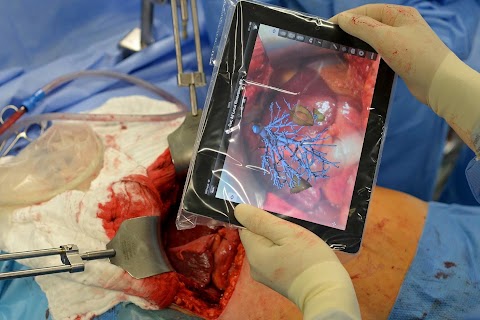
But, nevertheless, this photo taken at a hospital in Hamburg shows exactly that - a liver operation being performed with the support of a tablet computer. The device was used to access and visualise planning data, in one of the first surgeries of its kind in Germany.

Professor Oldhafer, chief physician of general and visceral surgery at Asklepios Hospital poses before the operation using the tablet computer.
The tablet uses augmented reality, which allows the liver to be filmed and overlaid during an operation with virtual 3D models, reconstructed from the real organ.
Developed by Fraunhofer MEVIS in Bremen, this procedure helps locate critical structures such as tumours and vessels, and is expected to make it easier to transfer plans made before the operation into actual surgery.
Slideshow

A co-worker ties Professor Karl Oldhafer's surgical gown before he goes into surgery.

He places a tablet computer into a sterile bag.

A doctor cranes his neck to observe the surgeons at work.

Professor Oldhafer poses with the tablet mid-operation.

He adjusts a digital image of the liver and its internal structures, seen next to the real organ.

A piece of liver is removed.

Professor Oldhafer exposes a tumour-ridden section of the liver.

Various surgical implements are used to sew up the patient's liver.

A piece of the tumorous liver lies next to a pair of scissors.
"Would I faint? Would I be horrified when they cut open the stomach with a long knife?"
When my boss, Joachim Herrmann, told me that I had to cover liver surgery using an iPad, I had no idea how an iPad could be helpful during an operation. I knew that iPhones, iPads and tablets were becoming more useful for all sorts of activities in our daily life – but for surgeries?
We use these new toys in many different ways, including GPS for cars, during sporting activities, music and mail. Some of my colleagues use tablet computers to present their portfolios and to operate their cameras. Swiss camera maker Alpa uses an iPhone as a viewfinder for their tilt and shift cameras. But I couldn’t imagine how an iPad would be helpful during an operation to remove two tumours from a liver.
Also, I knew nothing at all about livers or any surgery before this assignment.
To get a feel for the atmosphere, the hospital, light conditions and the team, I went to meet Professor Karl Oldhafer, chief physician of general and visceral surgery at the Asklepios Clinique in Hamburg-Barmbek, two days before I had to go through with my project. I felt slightly uncomfortable, being actually present at a surgical procedure. As I arrived at the hospital, I was confronted with an invitation by Professor Oldhafer to participate in a surgery there and then.
Never having seen this amount of blood, the massive cuts and all the different instruments being used, I was a little bit frightened. Would I faint? Would I be horrified when they cut open the stomach with a long knife? However, the “test run” went well. I survived and I had an idea what to expect in two days time.
After two more nights I woke early, had my usual pot of coffee and arrived pretty much on time at the Barmbek hospital to join Professor Oldhafer’s team in the changing room. The green clothing did fit well, but it wasn’t really my colour and also not my type of uniform.
The preparation was similar to the one I saw on the previous day. Then the Professor entered the room with an iPad in his hands. It was covered in a plastic bag, like the zip lock bags I use at home in my freezer.
When he could see the liver he used the iPad to locate the two tumours in it. It was very exciting as it was one of the first operations to be carried out in this way in Germany.
The tablet uses augmented reality, which allows the liver to be filmed with an iPad and overlaid during the operation, with virtual 3D models reconstructed from the real organ.
I was really impressed with how many surgeons, doctors and nurses took part in one surgery and how everybody seemed to know their roles exactly. Even more impressive and fascinating was getting a picture of this tiny little white lump – the cancer – which could be so vicious and dangerous to the human body.
After three hours of hard work by the whole team, myself included, the operation finished successfully. A great job was done and I want to thank Professor Oldhafer as well as his team for their kind support and especially Bianka Hofmann from the Fraunhofer MEWIS institute for her help and patience. Last but not least, a special thank you to Joachim in the Berlin office, who had the idea for this impressive story.
